


Clinical Case in Implant Dentistry Three-Year Evaluation by Dr. Carmy M. Michael DDS
Featured products
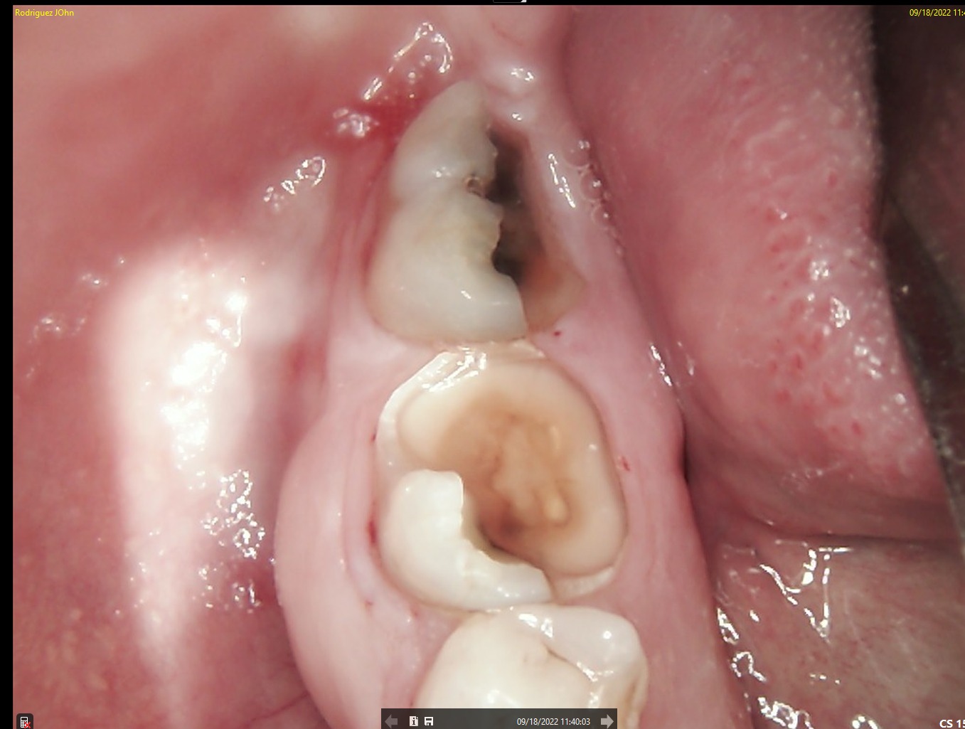
A patient presented with functional impairment and partial tooth loss in the posterior mandible. The main complaints included reduced chewing efficiency, and the need for a long-term, biologically stable restoration.
.jpeg)
.jpeg)
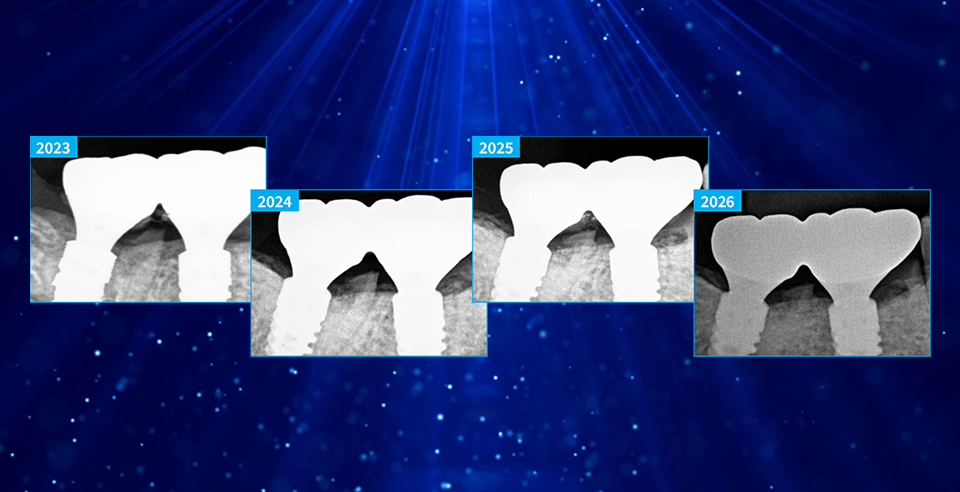
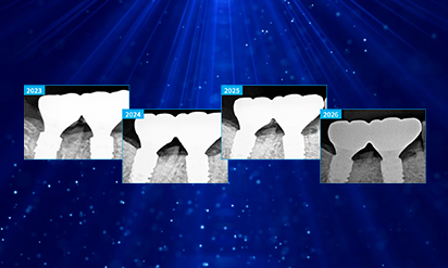
After comprehensive clinical and radiographic evaluation, a treatment plan was established to restore function and preserve peri-implant tissues using DSI Premium Implant System and a fully digital workflow. The case was documented from implant placement in 2023 through annual followups in 2024, 2025, and 2026, with a specific focus on the dynamic changes of peri-implant bone and soft tissue levels over a 3-year period.
INITIAL STATE
CBCT imaging was used for precise implant planning, including evaluation of bone volume and anatomical landmarks. Digital planning allowed optimal implant positioning to ensure long-term stability and prosthetic-driven placement. Intraoral and periapical images confirmed the clinical condition prior to surgical intervention.
.jpeg)
.jpeg)
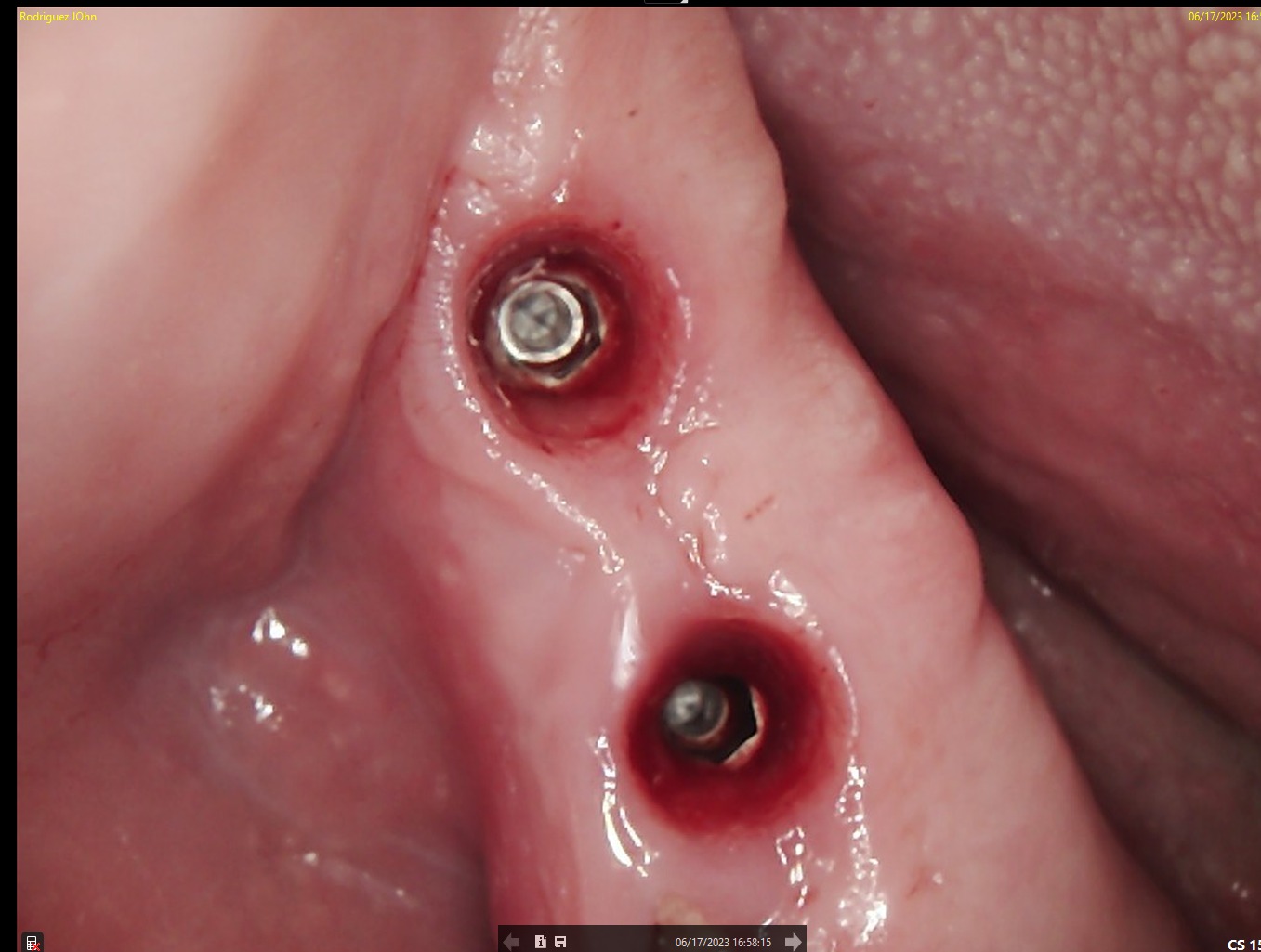
SURGERY
A flap approach was performed under local anesthesia. Osteotomies were prepared following the digital treatment plan, respecting biological principles and anatomical limitations. Two DSI Premium Mountless Implants were placed with high primary stability. The implants were positioned to allow optimal emergence profile and long term crestal bone preservation. Primary closure was achieved, and DSI Concave Healing Caps were placed to support soft tissue architecture during healing.
.jpeg)
HEALING STAGE
After the initial healing period:
• Soft tissue maturation was observed around the healing caps
• No signs of inflammation or peri-implant complications were noted
• Favorable gingival contour development was achieved
The concave design of the healing caps contributed to the formation of a stable peri-implant soft tissue seal.
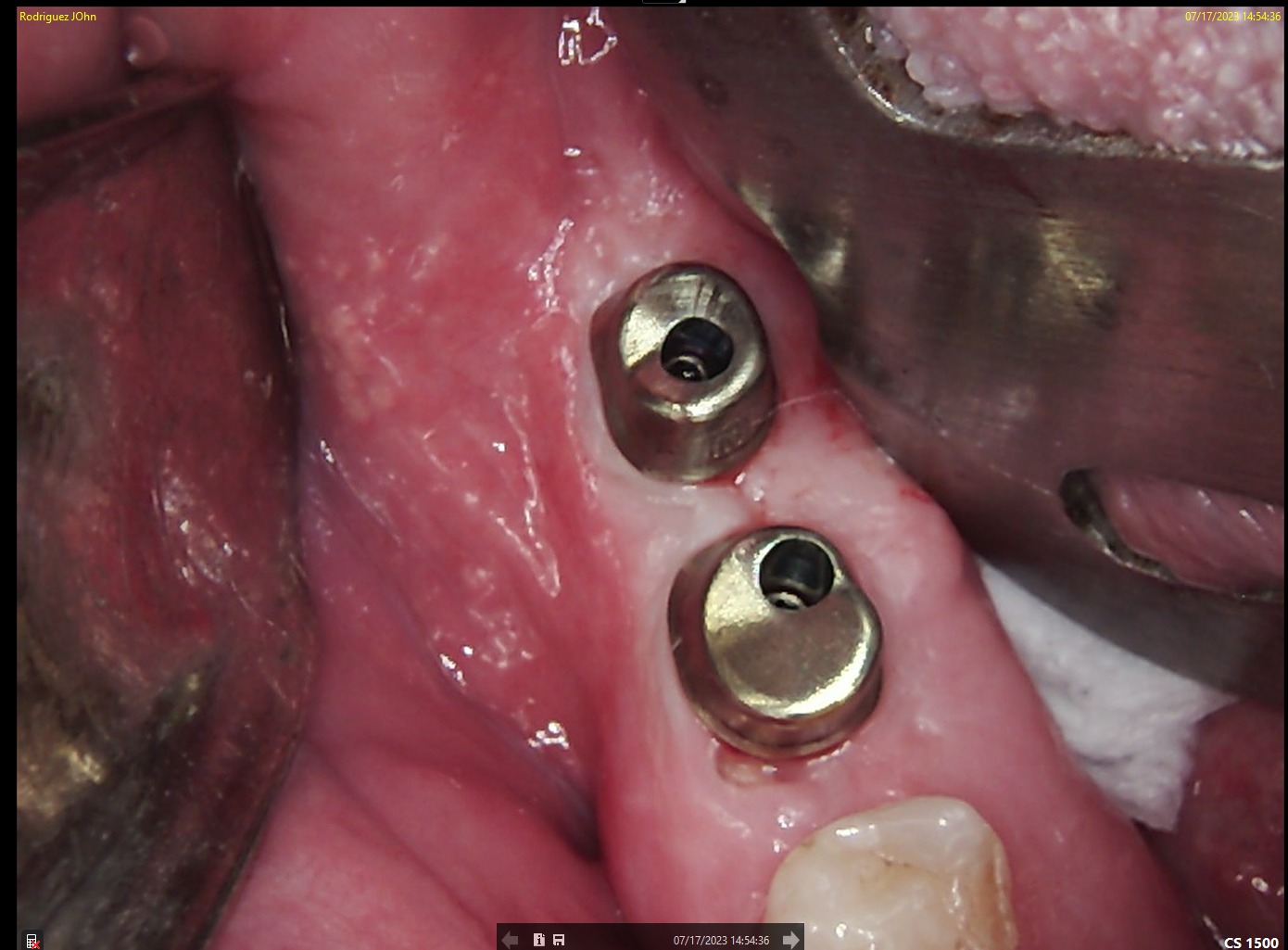
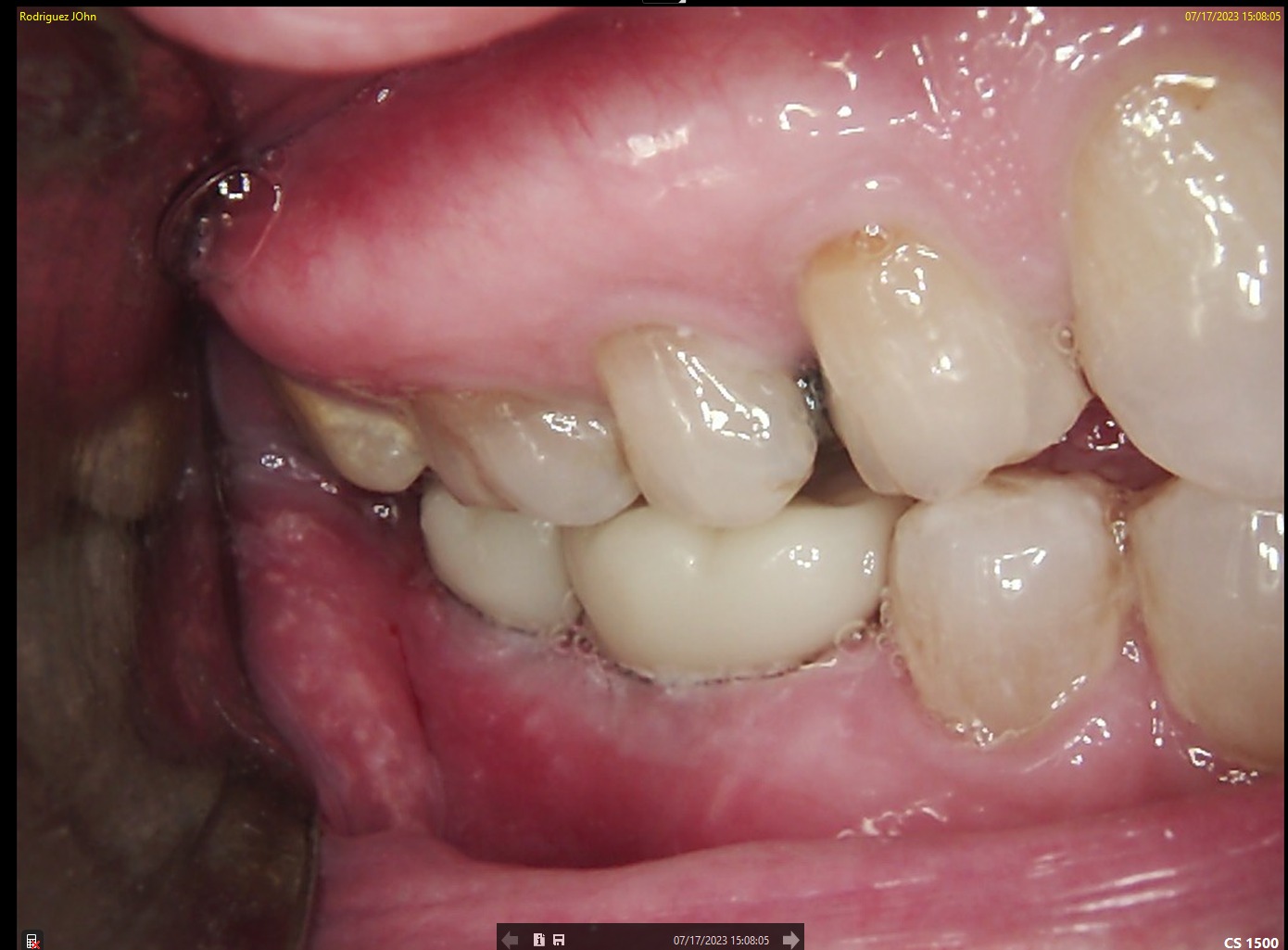
FINAL RESTORATION
Following successful osseointegration:
• Digital impressions were taken
• Screw-retained implant-supported restorations were fabricated using custom abutments
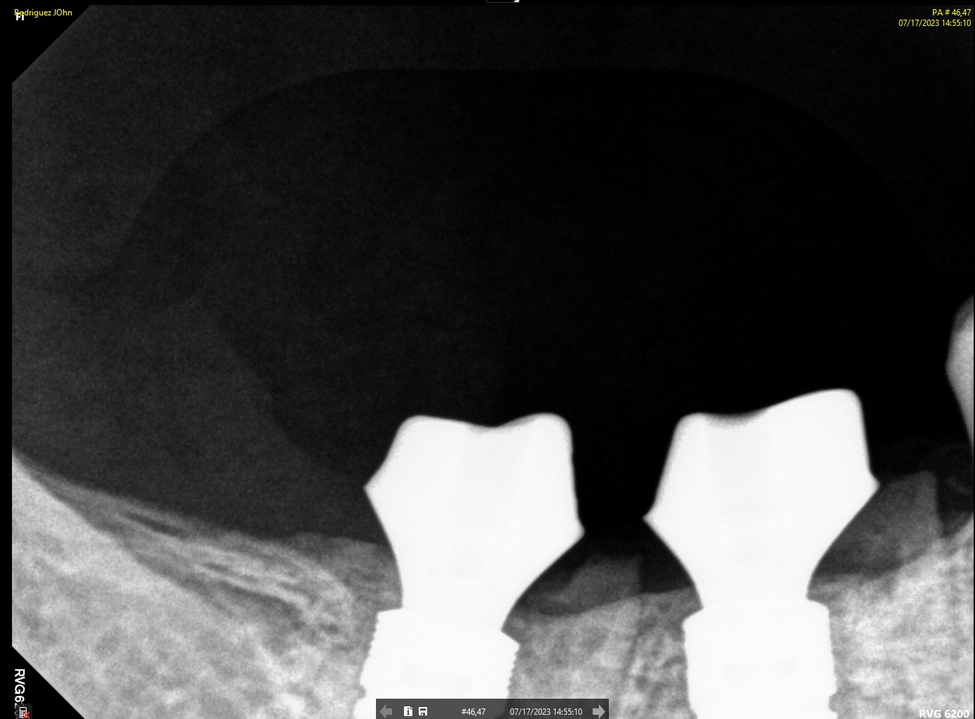
Final restorations demonstrated:
• Proper occlusal integration
• Natural emergence profile
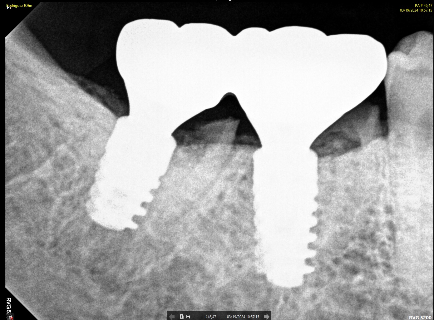
• Radiographic confirmation of stable crestal bone levels
Clinical and radiographic images confirmed successful functional and esthetic outcomes at delivery.
.jpeg)
.jpeg)
FOLLOW-UP - 2024
One-year follow-up evaluation showed:
• Stable peri-implant soft tissue contours
• Absence of inflammation or recession
• Radiographically preserved crestal bone levels
The implants remained fully functional with excellent tissue response.
.jpeg)
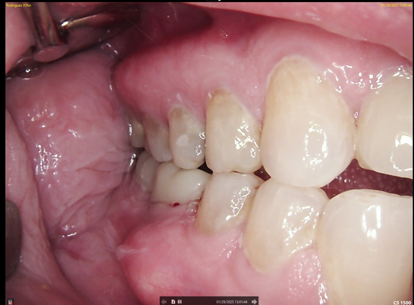
FOLLOW-UP - 2025
At the two-year follow-up:
• Continued soft tissue stability was observed
• No signs of peri-implant bone loss
• Prosthetic components remained intact and functional
Radiographic analysis confirmed long-term osseointegration and biological stability.
.jpeg)
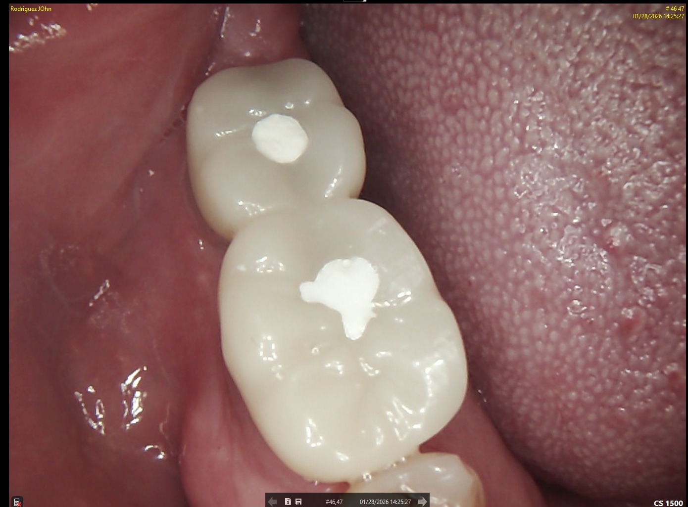
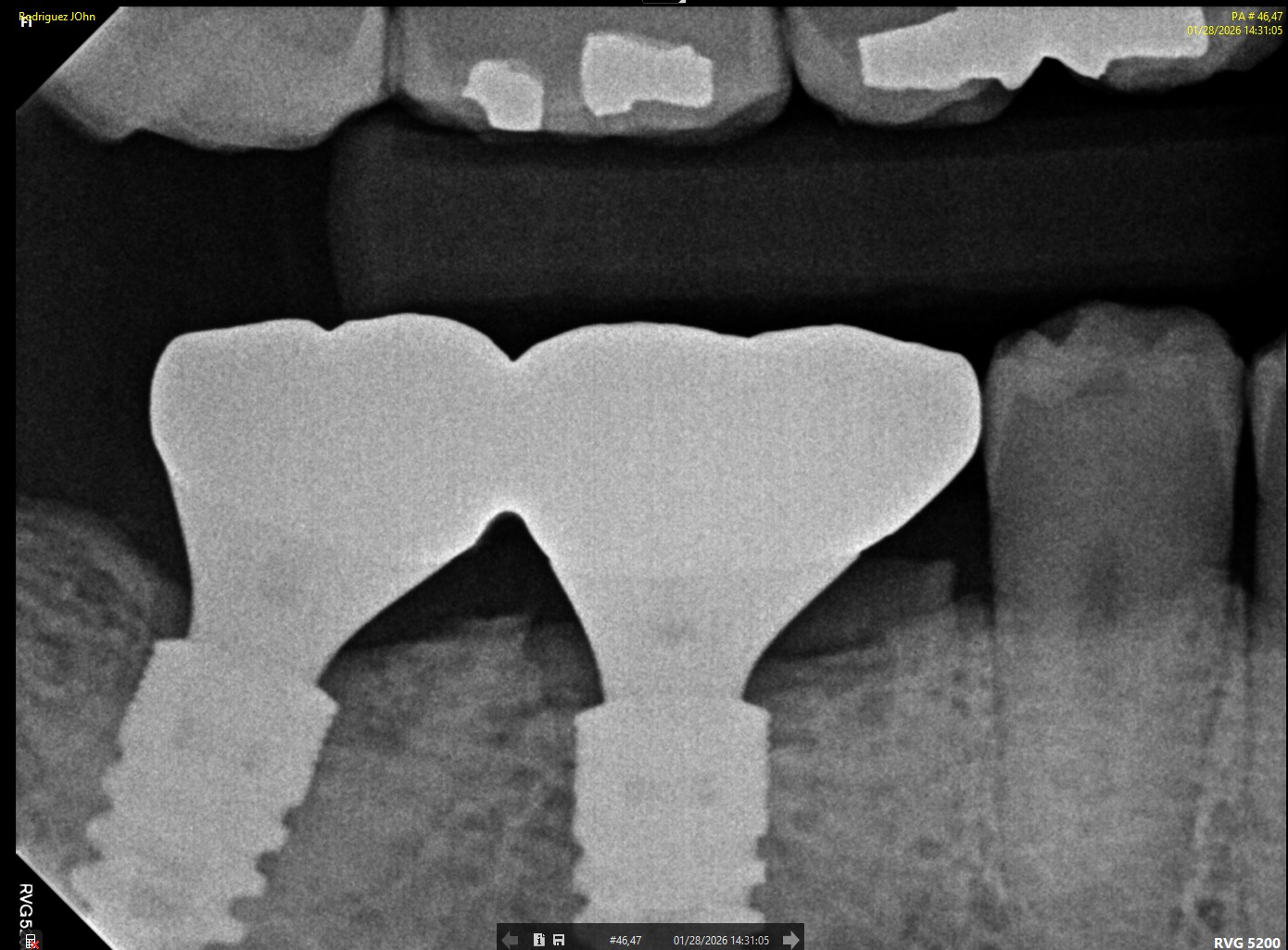
FOLLOW-UP - 2026
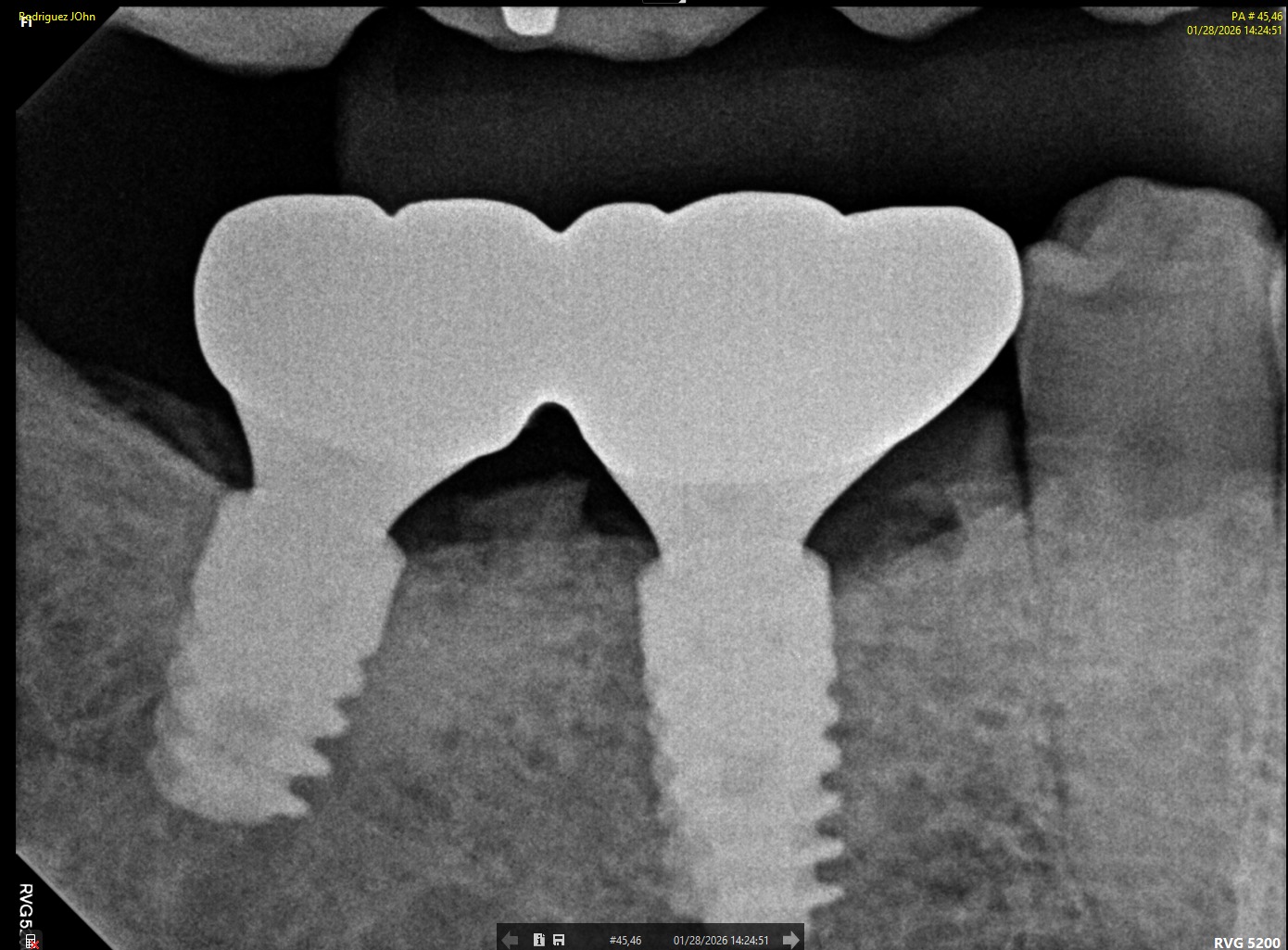
At the three-year follow-up:
• Peri-implant bone levels remained stable
• Soft tissues demonstrated healthy morphology and color
• The restorations maintained functional integrity under occlusal load
Both clinical and radiographic evaluations confirmed long-term hard and soft tissue stability, validating the treatment protocol and implant system used.
.jpeg)
CONCLUSION
This three-year clinical follow-up demonstrates that implant-supported restorations using DSI Dental Implants provide predictable and stable outcomes. The combination of accurate digital planning, biologically driven surgical techniques, and appropriate prosthetic components resulted in excellent preservation of peri-implant hard and soft tissues over time.